Regenyal Laboratories
Vai all'Italiano
Login / Register
  • About
  • Biorivolumetria
    • Green Injections & Technologies
    • Certifications
    • International Patent
    • Bio-Process
  • Products
    • CATEGORIES
      • Biorivolumetria
      • Multifractional BIO-INDUCTION
      • Intra-articular
      • Biorevitalizing creams
      • Medical device for topical use
    • BRAND
      • Regenyal Idea Bio-Expander
      • Regenyal Idea
      • Regenyal Idea Lips
      • Regenyal Super Idea KIT
      • BIOREGEN
      • Regenflex / Regenflex Starter / Regenflex BIO-PLUS
      • Regenyal face cream
      • Regenyal body cream
      • Regenyal sun care
      • Dermo Q.ore
    • FOR SPECIALISTS
      • Regenyal Idea Bio-Expander
      • Regenyal Idea
      • Regenyal Idea Lips
      • Regenyal Super Idea KIT
      • BIOREGEN
      • Regenflex / Regenflex Starter / Regenflex BIO-PLUS
      • Regenflex BIO-PLUS
  • PRE-POST
  • Press
    • Events
  • Video/Brochure
  • Contact
    • Partnership
+39 0735 757947

Regenflex / Regenflex Starter / Regenflex BIO-PLUS

Home / Shop / Regenflex / Regenflex / Regenflex Starter / Regenflex BIO-PLUS

REGENFLEX STARTER 1 syringe containing 2 ml of sterile pyrogen-free solution – (Hyaluronic acid sodium salt 1,6%)
REGENFLEX 1 syringe containing 2 ml of sterile pyrogen-free solution – (Hyaluronic acid sodium salt 0,8%)
REGENFLEX BIO-PLUS 1 pre-filled mono-use syringe (75 mg of reticulated hyaluronic acid in 3 ml buffered physiological solution).
Category: Regenflex, Intrarticolari.
  • Description
  • Hyaluronic Acid & Joints
  • Studies
  • Injection technique
  • Video

Regenflex / Regenflex Starter / Regenflex BIO-PLUS

REGENFLEX® is a medical device based on a highly-purified Hyaluronic Acid, with molecular weight between 800-1.200 kDalton.
Sodium hyaluronate is a fundamental component of the synovial fluid, as it is responsible of the visco-elastic properties of the latter.
The sodium hyaluronate contained in REGENFLEX® is obtained by means of fermentation, avoiding any kind of chemical modification treatment, in order to guarantee an absolutely pure product, free of remains alien to the physiological context of implementation.

REGENFLEX® STARTER and REGENFLEX® are injectable products based on a buffered physiological solution (respectively 1,6% and 0,8%) of hyaluronic acid sodium salt with significant viscoelastic properties.
The packages include one single-use syringe, containing 2ml of sterile apyrogen solution.

REGENFLEX STARTER: MAIN PROPERTIES

Regenflex: main properties

Cross-linked & linear intercalated Hyaluronic Acid

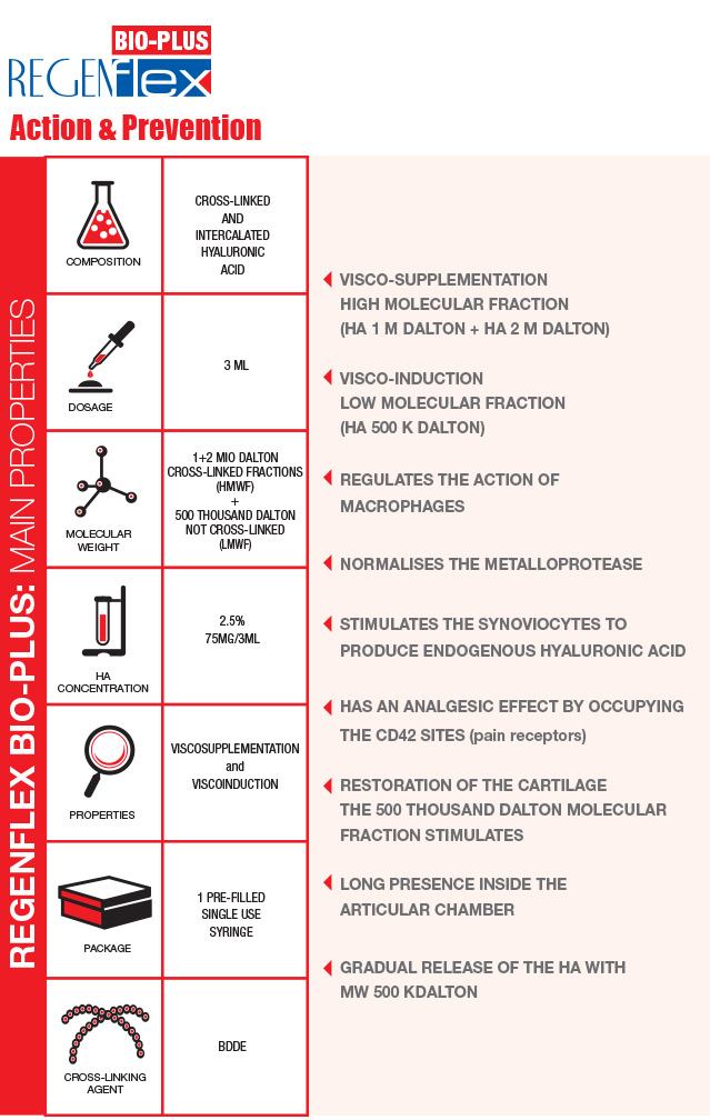
Regenflex BIO-PLUS is a transparent gel based on cross-linked and linear intercalated Hyaluronic acid, created with an advanced and patented technological process.
The aim of this intra-articular injectable is to prevent or to reduce pain, restoring the normal damping and lubricating function of the joint.
Not only will the joint function improve, but the patient will also experience an improvement in their quality of life.

The matter of weight: 2 molecular fractions

The action of the Hyaluronic acid depends on its molecular weight. The weight, in fact, influences the quantity of exogenous hyaluronic acid that can penetrate the synovial membrane: such quantity is proportionally inverse to the molecular weight itself. The close combination of the cross-linked and the intercalated linear Hyaluronic acid, makes it possible to inject two molecular fractions:

Regenflex BIO-PLUS & Biorivolumetria

BIORIVOLUMETRIA is an innovative concept in the field of HYALURONIC ACID-based injectiable medical devices.

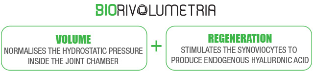
The same product makes it possible to simultaneously: restore the volume of the joint chamber by injecting CROSS-LINKED HYALURONIC ACID, and to stimulate receptors of the synoviocytes and chondrocytes thanks to the gradually released linear fraction of the INTERCALATED HA.

Manufacturing technology and functioning

The manufacturing technology used to produce Regenflex BIO-PLUS is the one used for BIORIVOLUMETRIA. The Biorivolumetria products are MONO-PHASE GELS obtained through a long-lasting cross-linking process at low temperature, the “gentle cross-linking”. The slow mixing allows for the cross-linking agent (BDDE) to widespread in a better and more uniform way. The amount of BDDE used is 30 per cent lower than in the majority of other products with equal degree of viscosity currently on the market. For the manufacturing of Regenflex BIO-PLUS three molecular weights of Hyaluronic Acid are used: 1 M, 2 M and 500 THOUSAND Dalton. The latter is linear and intercalated and represent 10 per cent of the total Hyaluronic Acid. Differently long chains of HA help to better calibrate the product’s viscosity. The linear Hyaluronic Acid is released gradually in time, performing a prolonged biological action (visco-induction).

Intercalation process

INTERVIEW TESTIMONIAL EMIDIO MORGANTI

brochure_regenflex_eng

What is Hyaluronic Acid (HA)

  • It is a natural component of the connective tissue
  • Given its ability to form connections with collagen, proteoglycans, fibronectin and water, it is the basis of large molecular units that support the extracellular matrix
  • It is an important component of the synovial fluid
  • Helps to lubricate the joint and to damp down mechanical stress (lubricating and damping function)
  • Protects the cartilage against the penetration of inflammatory cells and from the lytic enzymes

Hyaluronic Acid and Joints

In trauma-caused and degenerative diseases of the joints there is an in situ decrease of hyaluronic acid, due to which the synovial fluid is less viscous. Such deficiency determines a compression of the articular function and causes pain. THE INTRA-ARTICULAR ADMINISTRATION OF EXOGENOUS HYALURONIC ACID HELPS TO:

What risks are there for the joints?

The joints are subject to degenerative, sometimes chronic, diseases. The most common of these is the CHONDRAL DISEASE, that causes regressive alterations of:

Causes of the chondral disease

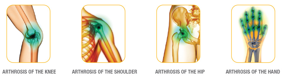
In the case of chondral diseases, the bones, which in normal conditions should slide against each other without friction because of their smooth and flexible tissue, are instead impaired in the movements. This is due to the loss of rheologic synovial properties, that causes a notable reduction of the synovial fluid’s visco-elasticity, leading to:

Study_1

A recent study on Regenflex Starter and Regenflex shows that using the products for at least three weeks offers the following results.
  • REDUCTION OF PAIN WITH NO JOINT LOADS
  • ATTENUATION OF PAIN WITH JOINT LOADS FROM THE FIRST ADMINISTRATION ON
  • IMPROVED SLIDING OF THE JOINT
  • INCREASED RANGE OF FLEXION AND EXTENSION OF THE JOINT
A statistically significant inprovement of the above values, compared with basic values, can be observed during the first six months of the use of REGENFLEX STARTER and REGENFLEX.

Study_2

Currently an observational study of 365 days is being conducted on 94 patients (43 males and 51 females) with 2nd degree by Kellgren-Lawrence chondral lesions of the knee (54 right knees and 40 left knees). Mean age of 69 years. No previous nor contemporary treatments with hyaluronic acid or chondroprotective compounds. The reparation/regeneration markers are assessed every three months with the ELISA TEST (Urinary C-terminal telopeptide of type II collagen CTX-II, Soluble receptors of Adiponectin, Visfatin and Leptin, Aggrecans), while the depth of the cartilage is evaluated by means of standard ultrasound scan. The ROM is measured with isokinetic tools, and the pain assessed against the VAS scale.
Results at 6 months
Significant increase of the progressive ROM with a proportional decrease of the pain. The ELISA TEST results will be published on completion of the study.

KNEE JOINT - FRONT INJECTION

  • Patient position Patient lying on the examination couch with head supported and knee bent approximately 90 degrees.
  • Reference point Patellar ligament, upper edge of lateral femoral condyle and upper edge of tibial plateau.
  • Injection point The point of least resistance between the lateral edge of patellar tendon and tibial plateau.
  • Needle angle Parallel to the tibial plateau, angled in the direction of the femoral groove.
  • Remarks The lateral meniscus is positioned close to the tibial plateau and should be avoided. This form of injection is useful in cases of patellofemoral arthritis. The entire needle length should penetrate the skin and should not encounter any resistance.

KNEE JOINT - SIDE INJECTION

  • Patient position Patient lying on the examination couch with head supported and knee extended.
  • Reference point Upper section of the patella and dorsal edge of lateral femoral condyle.
  • Injection point The point of least resistance between the upper section of the patella and dorsal edge of lateral femoral condyle.
  • Needle angle Parallel to the examination couch surface, angled in the direction of the patellar articular surface.
  • Remarks Useful to apply slight pressure on the medial side of the patella, which rotates and moves the patella laterally. In the event of intra-articular effusion, penetration should be carried out at the point of major swelling created by movement to the patella. The entire needle length should penetrate the skin and should not encounter any resistance. Should the patient experience pain, the point of the needle might well be on the synovial point. In such cases, the direction of the needle should be altered slightly until the pain is no longer felt.

ANKLE JOINT

  • Patient position Patient lying on the examination couch with head supported, knee extended and ankle in a neutral position.
  • Reference point The anterior tibial flexor tendon and anterior edge of the tibio-astralgic joint space.
  • Injection point Average proximal point between the junction of the anterior tibial tendon and tibio-astralgic joint space.
  • Needle angle Parallel to the articular face of the tibia, angled in the direction of the postero-lateral edge of the joint.
  • Remarks Pronosupination movement of the foot is useful in order to identify the point of least resistance at the medial side of the anterior tibial tendon. The entire needle length should penetrate the skin and should not encounter any resistance. Should the patient experience pain, the needle tip might well have punctured the synovial tissue. In such cases, change the direction of the needle slightly until the pain eases.

LOWER ASTRAGALIC JOINT

  • Patient position Patient lying on the examination couch with head supported, knee extended, the lower limb slightly intra-rotated and foot slightly arched.
  • Reference point Tarsal sinus.
  • Injection point Lower proximal corner of the tarsal sinus.
  • Needle angle Parallel to the foot arch surface, in the direction of the rear medial corner of the joint space.
  • Remarks The tarsal sinus is easily strengthened with the foot in supination position and joint space of the lower astragalic joint is easily strengthened where there is varus and valgus deformity on the heel bone.

TIBIAL TARSAL JOINT

  • Patient position Patient lying on the examination couch with head supported, knee extended, the lower limb slightly intra-rotated and foot slightly arched.
  • Reference point Tarsal sinus.
  • Injection point Point of least resistance between the lateral edge of the patellar tendon and tibial plateau.
  • Needle angle Parallel to the foot arch surface, in the direction of the rear medial corner of the joint space.
  • Remarks The tarsal sinus is easily strengthened with the foot in supination position and joint space of the lower astragalic joint is easily strengthened where there is varus and valgus deformity on the heel bone.

TRAPEZIOD-METACARPAL JOINT

  • Patient position Sat on the examination couch with knee bent at an angle of 90 degrees and arm slightly pronated.
  • Reference point Base of the first metacarpal bone.
  • Injection point In proximity to the base of the dorsal side face of the first metacarpal bone at approximately the centre of the joint.
  • Needle angle Perpendicular to the articular surface, angled in the direction of the centre of the joint.
  • Remarks Flex and extend the arm to aid identification of the joint space. It is worthwhile applying slight abduction to the hand and position the thumb slightly under traction.

HUMERO-RADIAL JOINT

  • Patient position Sat on the examination couch with knee bent at an angle of 90 degrees and forearm pronated.
  • Reference point Epicondyle.
  • Injection point Slightly higher then the epicondyle.
  • Needle angle Angled downwards at an angle of 30 degrees.
  • Remarks Medication is distributed along an arch of approximately 180m degrees, both above the finger extensor tendon and the short radial extensor muscle of the wrist, and below at the height of the epicondylar periosteum.

SUBACROMIAL SPACE

  • Patient position Seated.
  • Reference point Lower postero-lateral edge of the acromion.
  • Injection point The initial point of acromion anterior curvature.
  • Needle angle Angled slightly upwards in the direction of the centre of the space.
  • RemarksThe point of least resistance should be used. Recommended when pain is primarily due to tendon-muscle palpation of the supraspinous ligament.

HIP JOINT

  • Patient position Patient at rest, lying to one side and on the side opposite to that of the injection, with hips and knees bent at an approximate 40-degree angle.
  • Reference point At the emergence of the great trochanter.
  • Injection point Along the imaginary extension from the anterior inferior Iliac spine.
  • Needle angle The needle should be inserted at a right angle to the base support surface.
  • RemarksThe top of the great trochanter is normally immediately positioned under the cutaneous profile (with the exception of patients with severe adiposis) and it might well be necessary to withdraw the needle a few millimetres once the surface of the bone has been reached to avoid any significant resistance to the injection.

TEMPORO-MANDIBULAR JOINT

  • Patient position Seated with the head slightly rotated.
  • Reference point Mandibular condyle, mandibular pit of the temporal bone.
  • Injection point In front of the eye, below the zygomatic process in the mandibular pit of the temporal bone.
  • Needle angle Angled slightly upwards.
  • Remarks Ask the patient to open and close the mouth in order to identify a depression formed in the articular seat.
  • Filter by categories
    • Biorivolumetria
    • Multifractional BIO-INDUCTION
    • Intra-articular
    • Medical devices for topical use
    • Biorevitalizing creams
  • Filter by brand
    • Regenyal Idea Bio-Expander
    • Regenyal Idea
    • Regenyal Idea Lips
    • Regenyal Super Idea KIT
    • BIOREGEN
    • Regenflex / Regenflex Starter / Regenflex BIO-PLUS
    • Dermo Q.ore
    • Regenyal face cream
    • Regenyal body cream
    • Regenyal sun care
  • For specialists
    • Regenyal Idea Bio-Expander
    • Regenyal Idea
    • Regenyal Idea Lips
    • Regenyal Super Idea KIT
    • BIOREGEN
    • Regenflex / Regenflex Starter / Regenflex BIO-PLUS

Legal Address

  • San Benedetto del Tronto (AP) 63074, Via Valtellina, 21
  • Tel. +39 0735 757947
  • Fax. +39 0735 652039
  • esteri@regenyal.eu

Press

AMWC 2022

We are delighted to be a sponsor at the AMWC 2022 and we look forward to seeing you all there!

Continua...
AGORÀ 2021

AGORÀ 2021 from 14 to 16 October, MiCo-Milano Convention Center. Come and visit us: STAND A24

Continua...

REGENYAL LABORATORIES | C.F e P.I. 01942840446 |

Products | Privacy Policy | Certifications | Contact

Cambia lingua:
ITA | ENG

Select language

Regenyal Neutral Web Site

Regenyal Laboratories azienda cerificata